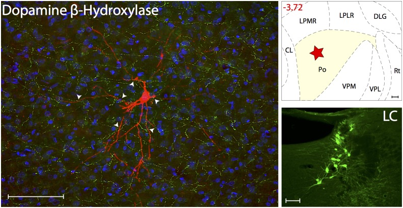
Figure 6. Noradrenergic innervation of thalamic trigeminovascular neurons.
Left: Immunopositive Dopamine β-Hydroxylase axons (green) surrounding a thalamic dura-sensitive neuron (red) labeled with TMR–dextran. Nuclear counterstaining was performed with DAPI (blue). Arrowheads indicate close apposition of DBH positive axons and the cell body and dendrites of the labeled neuron. Upper right: Location of the dura-sensitive neuron (red star) shown at left. Number in red indicates distance from bregma (mm). Lower right: Fluorescent image showing DBH labeling of cell bodies in the locus coeruleus (LC) of the brainstem. Scale bars = 100 µm.

