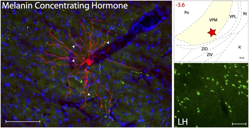
Figure 9. MCH innervation of thalamic trigeminovascular neurons.
Left: Immunopositive Melanin Concentrating Hormone axons (green) surrounding a thalamic dura-sensitive neuron (red) labeled with TMR–dextran. Nuclear counterstaining was performed with DAPI (blue). Arrowheads indicate close apposition of MCH positive axons and the dendrites of the labeled neuron. Upper right: Location of the dura-sensitive neuron (red star) shown at left. Number in red indicates distance from bregma (mm). Lower right: Fluorescent image showing MCH labeling of cell bodies in the lateral hypothalamus (LH). Scale bars = 100 µm. Abbreviations: ZID, zona incerta, dorsal; ZIV, zona incerta, ventral.

