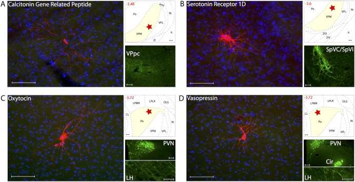
Figure 11. Lack of innervation of thalamic trigeminovascular neurons by axons containing CGRP, Serotonin 1D receptor, Oxytocin and Vasopressin.
Left A–D: Thalamic dura-sensitive neurons (red) labeled with TMR–dextran and nuclear counterstain with DAPI (blue). Note the absence of axonal immunoreactivity to CGRP (A), Serotonin 1D receptor (B), Oxytocin (C) and Vasopressin (D). Upper right A–D: Locations of the dura-sensitive neurons (red stars) shown at left. Numbers in red indicate distance from bregma (mm). Lower right A–B: Fluorescent images showing CGRP (A) and Serotonin 1D receptor (B) immunopositive axons in the parvicellular division of the ventral posterior thalamic nucleus (VPpc) and the spinal trigeminal nuclei (SpVC/SpVI; caudal/interpolar), respectively. Lower right C: Fluorescent images showing Oxytocin labeling of cell bodies and axons in the hypothalamic paraventricular nucleus (PVN) and lateral hypothalamus (LH), respectively. Lower right D: Fluorescent images showing Vasopressin labeling of cell bodies in the PVN and circular (Cir) nuclei of the hypothalamus, and axons in the LH. Scale bars = 100 µm.

