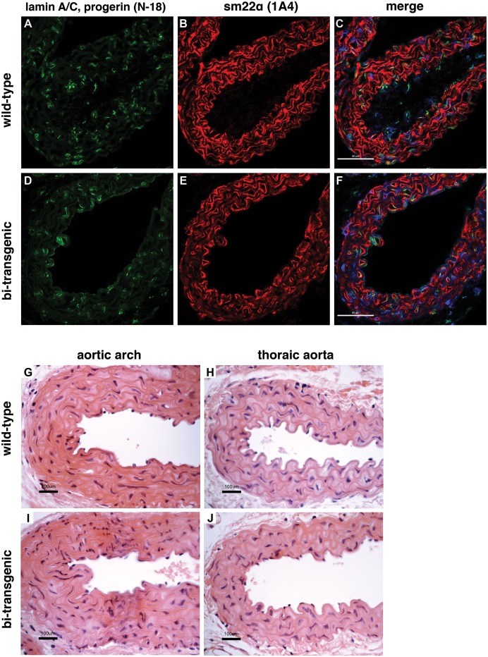
Figure 2. Expression of mouse lamin A/C and sm22α-actin is unaffected in the aortic arch.
Immunofluorescence staining with an anti-human-lamin A/C antibody (N-18), which also binds to progerin of human origin and lamin A/C of mouse origin, and an antibody for vascular smooth muscle cells (1A4) in wild-type (A–C) and bi-transgenic tetop-LAG608G+; sm22α-rtTA+ animals (D–F). Representative images from the sections of aortic arches of mice with the C57BL/6J; FVB/NCrl genetic background supplied with doxycycline from the date of birth until postnatal week 12. Scale bars: 50 µm. C, F: merge of the lamin A/C, sm22α-actin, and DAPI fluorescence signals. (G–J) Histological examination of aortic sections with haematoxylin eosin staining shows normal structure of the aorta. G, I: aortic arch. H, J: thoraic aorta. Scale bars: 100 µm.

