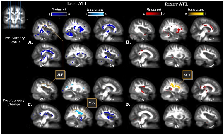
Figure 1. FA status in TLE patients before surgery.
Left TLE in the left (panel A) and right TLE in the right (panel B). Post-surgery changes are displayed for left (panel C) and right (panel D) ATL patients. Slice positions are displayed in the upper left corner. The six slices displayed in each panel are located in MNI space x = [−40 −27 −8 +8 +28 +40]. Abbreviations indicate SLF = superior longitudinal fasciculus, and SCR = superior corona radiata.

