
Figure 4.
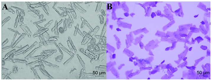
Isolated left ventricular myocytes of the rats with hypertension and heart failure (H-HF) at week 17 after clipping. (A) Low contrast bright field image (magnification, x200). (B) Hematoxylin and eosin whole cell staining (magnification, x200).