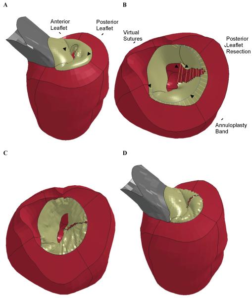
Figure 2.
Finite element simulation of triangular resection of P2 prolapse and annuloplasty. A) Pre-op model showing prolapse of the P2 leaflet. B) After triangular resection of the P2 section of the posterior leaflet. Virtual sutures can be seen connecting the cut edges of the posterior leaflet and between the annuloplasty band and the posterior annulus. The aortic root has been removed for clarity. C) Post-op model showing the mitral valve after virtual leaflet repair and annuloplasty in early diastole with the valve open. The aortic root has been removed for clarity. D) Post-op model at end-systole with the valve closed.

