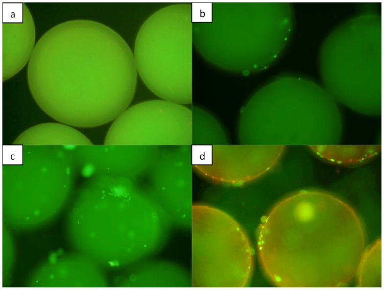
Fig 6.

Live/dead fluorescence image of microparticles a) without cells (control), b) without any growth factors on day 5, c) encapsulated with BMP-7, seeded with murine osteoblasts on day 5, d) without any growth factor on day 10. Microparticles are three dimensional and therefore in order to capture all the cells attached throughout the surface of the particle, different views were considered starting from periphery of the particle to its upper surface.
