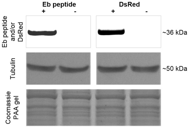
Figure 3.
Immunoblotting of hybrid dsRed-hEb protein. Sixty micrograms of protein were loaded in each lane. A 36 kD band can be detected in the lane where U2OS-dsRed-hEb extracts of stable cells is resolved and probed with anti-Eb antibody (panel 1 on the left). The same extract on a different PVDF membrane was probed with anti-dsRed antibody and a 36 kD band is visible (panel 1 on the right). No bands are evident in lanes containing proteins from naïve U2OS extracts. As a loading controls are shown, tubulin (~50 kD) and Coomassie blue staining (panel 2 and 3).

