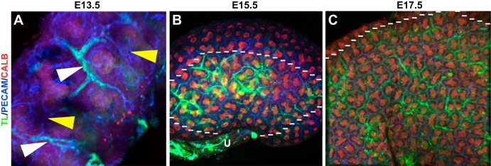
Fig. 2.

Whole mount images suggesting that the formation of renal vessels precedes renal blood flow. A–C: whole mount images of blood flow in the developing kidney (green) at various developmental stages colabeled with vascular [platelet endothelial cell adhesion molecule (PECAM; blue)] and ureteric epithelial (calbindin, red) markers. A: representative E13.5 kidney showing the major vessels (white arrowhead) that are perfused and peripheral areas of the kidney that are vascularized but lacking blood flow (yellow arrowheads). B: E15.5 whole mount kidney showing that the more centralized vessels (inside the dotted line) that are closer to the ureter (U) and angiogenic vessels are perfused and interdigitating between the ureteric epithelium (red), but the peripheral vessels (outside the dotted line), likely of vasculogenic origin, are unperfused. C: representative E17.5 kidney showing significantly more perfusion throughout the kidney interdigitating between the branching ureteric epithelium (below the dotted line). However, in the presumptive nephrogenic zone, the vessels are unperfused (above the dotted line).
