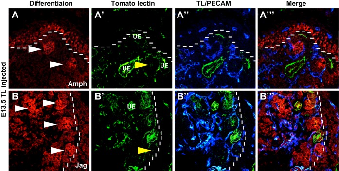
Fig. 6.
Renal blood flow is closely associated with nephron differentiation. A and B: representative images of E13.5 TL (green)-injected kidneys stained for nephron differentiation markers amphyphisin and jagged1 (Amph and Jag; red) and the vasculature (PECAM, blue). A–A″′: amphiphysin (red) staining was found in the early renal vesicles (white arrowheads) and also throughout the nephron progenitors in the neprogenic zone (dotted line). Perfused vessels were found encircling these differentiating structures and also contained within them (yellow arrowheads). B–B″′: similarly, jagged1-positive renal vesicles (white arrowheads) were observed surrounded by perfused vessels. The vessels were also located within the vesicles (yellow arrowheads).

