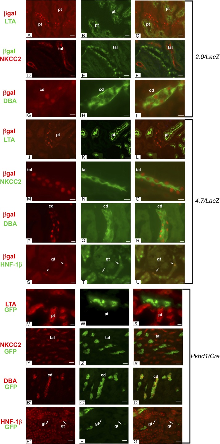
Fig. 4.
Expression of 2.0- and 4.7-kb transgenes in the kidney. A: antibody staining of kidney sections from adult 2.0/lacZ mice showed localization of E. coli β-galactosidase (red) in nuclei. B: costaining with FITC-conjugated Lotus tetragonolobus agglutinin (LTA; green) labeled proximal tubules (pt). C: merged image showing that the 2.0/lacZ transgene was not expressed in proximal tubules. D–F: costaining with antibodies against β-galactosidase (green) and Na+-K+-2Cl− cotransporter (NKCC2; red) showed no expression of the 2.0/lacZ transgene in thick ascending limbs of loops of Henle (tal). G–I: costaining with anti-β-galactosidase (red) and FITC-conjugated Dolichos biflorus agglutinin (DBA; green) showed expression of the 2.0/lacZ transgene in collecting ducts (cd). J–L: staining of kidney sections from adult 4.7/lacZ mice with an anti-β-galactosidase antibody (red) and FITC-conjugated LTA (green) showed that the transgene was not expressed in proximal tubules. M–O: costaining with antibodies against β-galactosidase (red) and NKCC2 (green) showed expression of the 4.7/lacZ transgene in thick ascending limbs of loops of Henle. P–R: costaining with anti-β-galactosidase antibody (red) and FITC-conjugated DBA (green) showed expression of the 4.7/lacZ transgene in collecting ducts. S–U: staining of the renal cortex from 4.7/lacZ mice with anti-β-galactosidase antibody (red) showed nuclear staining in parietal epithelia cells of Bowman's capsule (arrows), which also expressed HNF-1β (green). No transgene expression was observed in the glomerular tufts (gt). V–X: staining of kidney sections from adult Pkhd1/Cre;RYFP mice with an anti-green fluorescent protein (GFP) antibody (green) and biotinylated LTA followed by detection with rhodamine avidin D (red) showed that the transgene was not expressed in proximal tubules. Y–A′: costaining with antibodies against GFP (green) and NKCC2 (red) showed that the Pkhd1/Cre transgene was not expressed in thick ascending limbs of loops of Henle. B′–D′: costaining with anti-GFP antibody (green) and biotinylated DBA (red) showed expression of the Pkhd1/Cre transgene in collecting ducts. E′–G′: staining of the renal cortex from Pkhd1/Cre;RYFP mice with antibodies against GFP (green) and HNF-1β (red) showed no GFP expression in Bowman's capsule (arrows). Scale bars = 10 μm.

