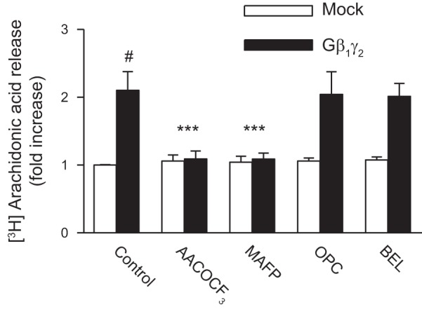
Fig. 7.

Effects of PLA2 inhibitors on AA release. Subconfluent proximal tubular cells were labeled with 0.5 μCi·ml−1·well−1 [3H]AA for 4 h before treatment. They were treated with10 μmol/l AACOCF3, MAFP, OPC, or BEL for 15 min or the vehicle DMSO, then transfected with Gβ1γ2 or empty vector (mock) for 24 h. At the end of incubation, an aliquot of medium was removed and counted for radioactivity. Values are means ± SE of 4 independent experiments shown as fold-increase. #P < 0.001, compared with nonstimulated control (mock) cells. ***P < 0.001, compared with the respective values of cells transfected with β1γ2 alone.
