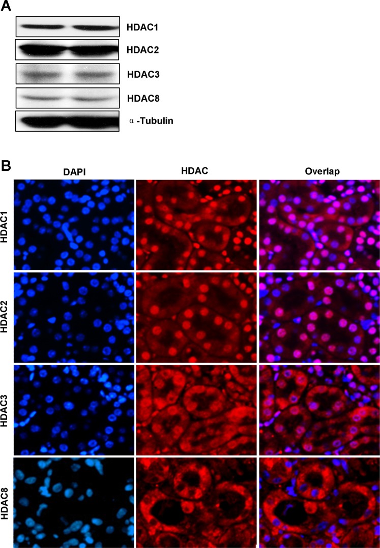
Fig. 1.
Expression and location of class I histone deacetylases (HDACs) in the kidney. A: tissue lysates from 2 normal kidneys were subjected to immunoblot analysis with specific antibodies against HDAC1, HDAC2, HDAC3, HDAC8, and α-tubulin. B: photomicrographs (×200) illustrate immunofluorescent costaining of HDAC1, HDAC2, HDAC3, or HDAC8 with DAPI in normal kidney sections.

