Fig. 3.
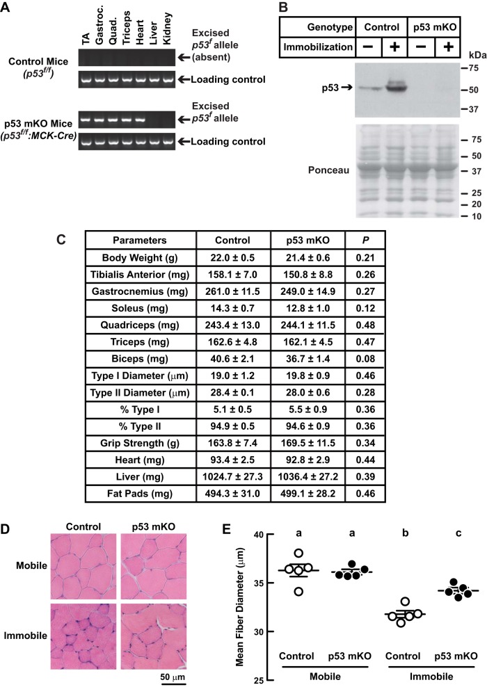
p53 is partially required for immobilization-induced muscle atrophy. A–E: p53-muscle knockout (mKO) mice are homozygous for a floxed p53 allele (p53f/f) and possess the MCK-Cre transgene, which directs Cre recombinase expression to skeletal muscle fibers and heart. Control mice were littermates of p53-mKO mice that lacked the MCK-Cre transgene. A: PCR confirmation that MCK-Cre directs excision of the floxed p53 allele (p53f) in striated muscle of p53-mKO mice. Gastroc, gastrocnemius; Quad, quadriceps. B: control and p53-mKO mice were subjected to unilateral hindlimb immobilization for 3 days before bilateral TA muscles were harvested. An equal amount of protein from each muscle (100 μg) was subjected to SDS-PAGE and immunoblot analysis with anti-p53 polyclonal IgG. Membranes were stained with Ponceau S to confirm equal loading. C: baseline analysis of control and p53-mKO mice. Data are means ± SE from 8 mice/genotype. Muscle weights are combined weight of bilateral muscles. Fat pad weights are combined weights of bilateral epididymal, retroperitoneal, and scapular fat pads. P values were determined with unpaired t-tests. D and E: control and p53-mKO mice were subjected to unilateral hindlimb immobilization for 3 days before bilateral TA muscles were harvested for histological analysis. Representative H & E images (D) and quantification of TA muscle fiber size (E). Each data point represents the mean diameter of ≥250 fibers from 1 muscle. Horizontal bars denote means ± SE. Statistical analysis used the 1-way ANOVA with Sidak's post hoc test; different letters are statistically different (P ≤ 0.05).

