Fig. 6.
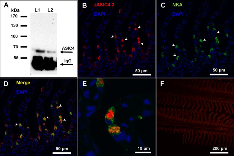
Immunoreactivity of anti-zebrafish ASIC4.2 (zASIC4.2) antibody in the gill of rainbow trout. A: immunoprecipitation Western blot from whole gill homogenates with zASIC4.2 showing a distinct band of ∼65 kDa. Lane 1 (L1) contains 300 mg of gill tissue, and lane 2 (L2) contains 200 mg of gill tissue. B–E: confocal images of gill sections labeled for ASIC4 and Na+-K+-ATPase (NKA) proteins. B and C: double-labeling with DAPI and anti-zASIC4.2 (B) and DAPI and anti-NKA (C). D: merged image of B and C. E: higher-magnification image of a MRC showing that ASIC4 and NKA are present in distinct regions within the MRC. F: control micrograph with DAPI and no primary antibody. Arrowheads (B–D) indicate positive staining.

