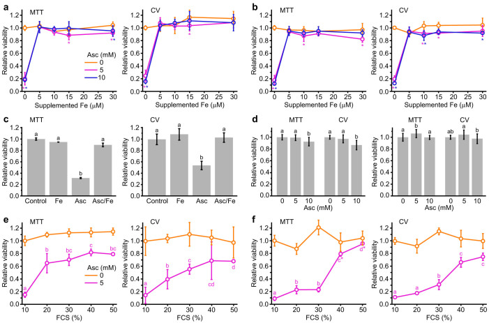
Figure 1. Effects of pharmacological ascorbate and physiological iron on the viability of cancer and primary cells according to MTT and CV assays.
(a) LNCaP and (b) PC-3 cells treated in cell culture medium (RPMI-1640 + 10% FCS) with different concentrations of supplemented iron; * - statistically significant (p < 0.05) compared to control with the same amount of supplemented iron. (c) Primary astrocytes treated with Asc (5 mM) and/or iron (30 μM) in cell culture medium. (d) LNCaP (left) and PC-3 (right) in human plasma; Bars not sharing a common letter are significantly different (p < 0.05). (e) LNCaP and (f) PC-3 cells treated in cell culture medium with different % (v/v) of FCS; Bars not sharing a common letter are significantly different (p < 0.05); * - non-significant compared to control with the same % (v/v) of FCS. Statistical analysis was performed using one-way or two-way ANOVA with post hoc Duncan test.

