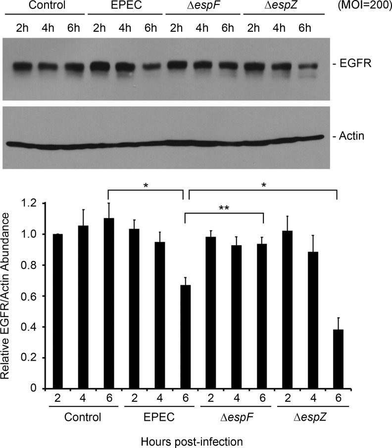
Fig. 6.
Validation of EPEC-mediated EGFR degradation in T84 cells. T84 cells were treated with medium alone or infected with EPEC, ΔespF mutant, or ΔespZ strain at MOI = 200. Total protein extracts isolated at specific time points were analyzed for the abundance of EGFR and actin by Western hybridization. Blots shown are representative of 3 similar experiments. Densitometric analysis was performed to determine EGFR abundance relative to actin and normalized to levels in control uninfected cells. Significant changes were assessed by ANOVA with Bonferroni post hoc test. *P < 0.01 and **P < 0.05 for specific conditions compared. Error bars represent standard error.

