Fig. 2.
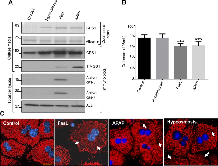
Primary hepatocytes release CPS1 in response to different types of liver injury. A: primary mouse hepatocytes were cultured in the presence of hypoosmotic medium (200 mosmol/l), FasL (0.5 μg/ml), and acetaminophen (APAP, 1 mM). The concentrated cell culture medium (25 μl) and total cell lysates were used for immunoblotting with antibodies to the indicated proteins. B: after each treatment, viable cell count was determined by Trypan blue staining after trypsinization. Each bar represents mean counts of 10–15 fields. ***P < 0.001 (compared with control cells). C: mouse hepatocytes were challenged with FasL, APAP, and hypoosmosis as in A. Cells were fixed and immunostained with CPS1 (red) and counterstained with DAPI (blue). Arrows highlight areas with less dense CPS1 staining. Scale bar = 20 μm. The displayed results were from a single mouse hepatocyte isolation with similar results obtained from 2 additional independent experiments.

