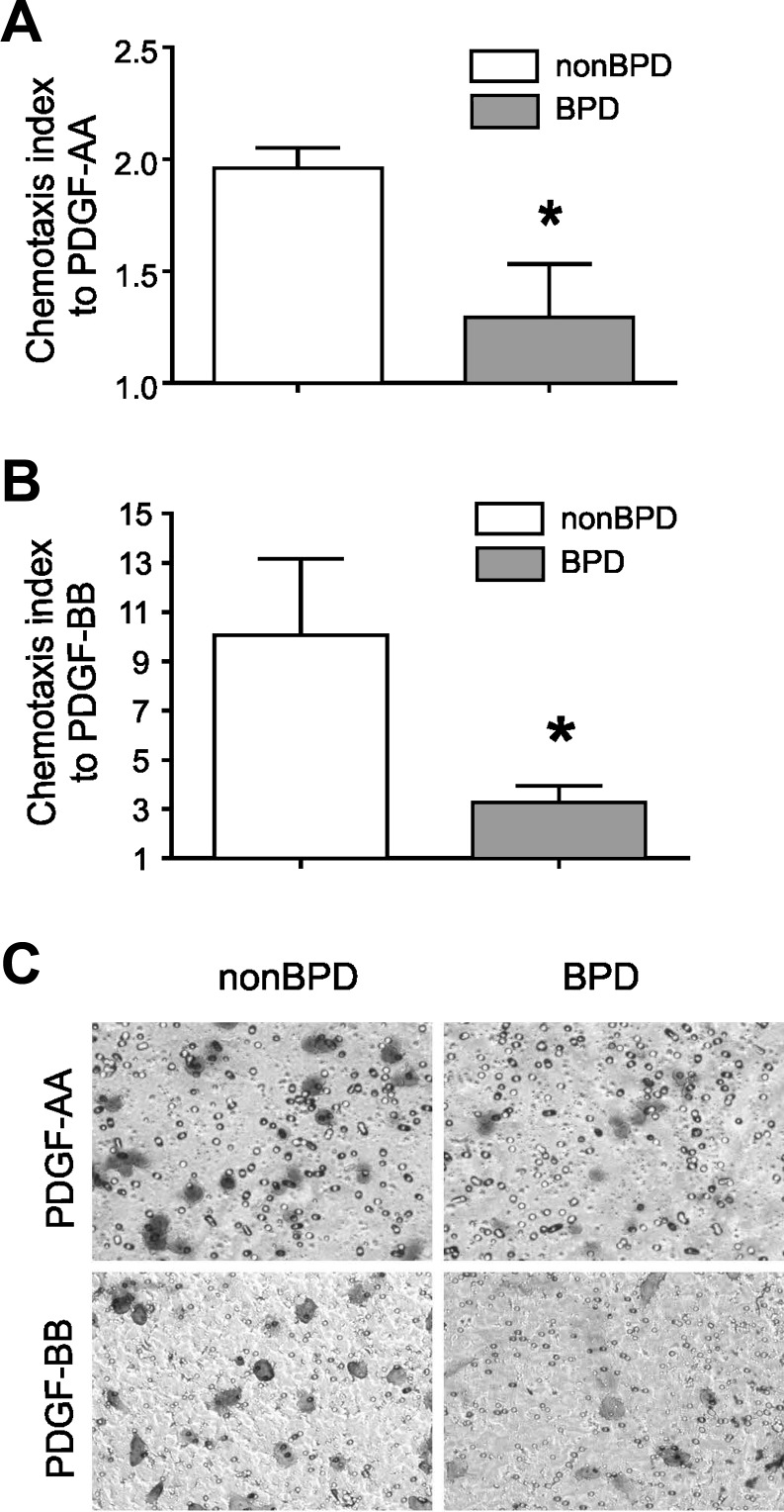
Fig. 3.
Neonatal lung MSCs from infants who develop BPD show decreased migration to PDGF. Migration of neonatal lung MSCs in response to PDGF-AA (30 ng/ml) and PDGF-BB (10 ng/ml) was assessed after 4-h incubation in 12-well Boyden chamber. PDGF-AA (A) and PDGF-BB (B). Group mean chemotaxis indexes for PDGF-AA (A) and PDGF-BB (B) are shown (*P < 0.05, unpaired t-test) compared with those for neonatal lung MSCs from infants not developing disease. In C, a representative membrane, stained after 4 h incubation with PDGF-AA or PDGF-BB, shows that fewer MSCs from infants who developed BPD cross through the membrane (toluidine blue stain). Results shown are representative of 3 independent experiments.

