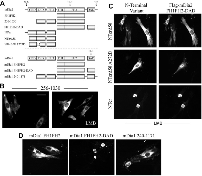
Fig. 6.
The FH2 NLS was masked by the autoinhibited state of mDia2. A: schematic of the mDia2 and mDia1 variants used in these experiments. B: localization of the mDia2 deletion variant 256–1030 in 10T1/2 cells in the absence and presence of LMB. Scale bar = 20 μm. C: FH1FH2-DAD localization was examined in LMB-treated 10T1/2 cells coexpressing the indicated NH2-terminal (NTer) mDia2 fragments. D: localization of the indicated mDia1 variants in 10T1/2 cells.

