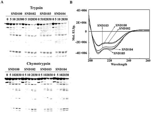
FIG. 7.
The structural properties of the N-terminally truncated σA. (A) Partial proteolysis analyses of the N-terminally truncated σA. The truncated σA subunits were partially digested with trypsin (top panel) or chymotrypsin (bottom panel) (see Materials and Methods). The time points (in minutes) to stop the proteolytic reaction are shown on top of each panel. (B) CD spectra of the N-terminally truncated σA. Each of the N-terminally truncated σA (0.2 mg/ml) subunits was scanned with UV light from 190 to 260 nm. The absorption curves for the truncated σA are as indicated.

