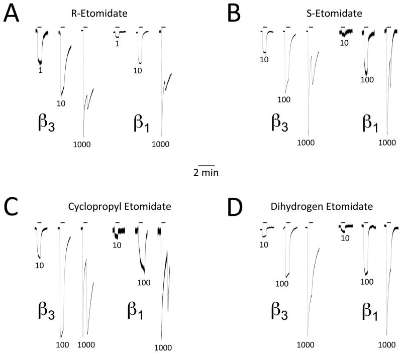
Figure 3.
Representative two-microelectrode electrophysiological traces recorded upon applying R-etomidate (A), S-etomidate (B), cyclopropyl etomidate (C), or dihydrogen etomidate (D) at the indicated concentrations (in μM) for 30 sec. Traces obtained from oocytes expressing α1(L264T)β3γ2L and α1(L264T)β1γ2L γ-aminobutyric acid type A receptors are labeled as β3 and β1, respectively. Each panel shows the effect of three different drug concentrations on currents mediated by the two receptor subtypes. In each set of three traces, the peak current amplitudes have been normalized to that produced by 100 μM γ-aminobutyric acid in the same oocyte.

