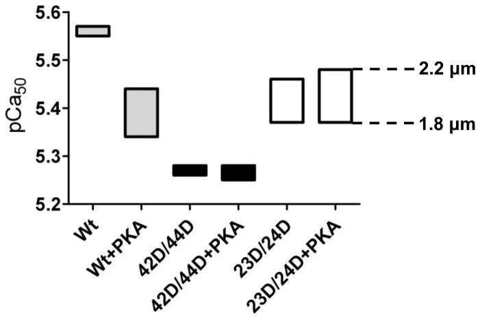
Figure 6. Schematic representation of changes in length-dependent Ca2+-sensitivity upon phosphorylation of Ser42/44 and/or Ser23/24.
Data obtained in troponin-exchanged donor cells without and with treatment with exogenous PKA (data from Figure 4 and Table 3 and 4) were combined to illustrate the range at which myofilament Ca2+-sensitivity (pCa50) may vary in response to phosphorylation at the PKC sites Ser42/44 and at Ser23/24. Abbreviations: wild-type (Wt); pseudo-phosphorylated 42/44 (42D/44D); pseudo-phosphorylated Ser23/24 (23D/24D). Boxes represent the range of Ca2+-sensitivity measured at a sarcomere length of 1.8 (bottom line) and 2.2 μm (upper line). This figure demonstrates that the sarcomere length-dependent shift in Ca2+-sensitivity is relatively small in Wt and 42D/44D without PKA (i.e. when cTnI phosphorylation at Ser23/24 is low). PKA treatment of Wt and Ser23/24 pseudo-phosphorylation enhance the length-dependent increase in Ca2+-sensitivity. However, PKA treatment of 42D/44D does not increase the Ca2+-sensitivity range at which the sarcomere is operating between sarcomere lengths of 1.8 and 2.2 μm.

