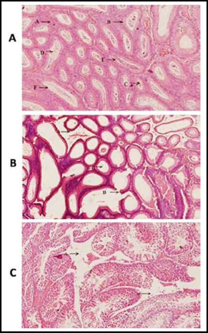
Fig.2.
a) Cross section of testicle of mice of control group at 60th day of experiment. A. Leydig Cells, B. Germinal Layer, C. Spermatids, D. Spermatocytes, E. Spermatogonia, F. Sertoli cell.
b) Cross section of testicle of mice of group B at 60th day. A. Destruction of Leydig cells and D. destruction of complete follicles.
c) Cross section of testicle of mice of group C at 60th day. A. Destruction of Leydig cells, B. Empty central lumen of follicle of testis without sperms, C. partially damaged germinal layer D. destruction of complete follicle.

