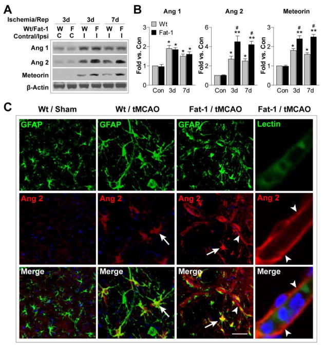
Figure 3. n-3 PUFA overproduction elevates angiopoietin 2 expression after tMCAO.
A. Representative Western blots of angiopoietin 1 (Ang 1), angiopoietin 2 (Ang 2), and meteorin in the contralateral and ipsilateral hemispheres of Wt and fat-1 brains after 3 or 7 d of reperfusion. β-actin was used as an internal loading control. B. Protein expression of Ang 1, Ang 2 and meteorin was quantified and expressed relative to the contralateral side (Con) of Wt mice. Data are presented as mean ± SEM, n=4–5 animals/group. *p≤0.05, **p≤0.01 vs. Con. #p≤0.05 fat-1 vs. Wt. C. Representative images of Ang 2 immunofluorescent signal (red), double labeled with GFAP or lectin (green), in the IBZ of Wt and fat-1 brains 7 d after tMCAO. Sections were counterstained with DAPI (blue) for nuclear labeling. Arrow: Increased Ang 2 expression in GFAP+ astrocytes. Arrowhead: Enhanced Ang 2 signal along the microvessels. Scale bar: 20 μm.

