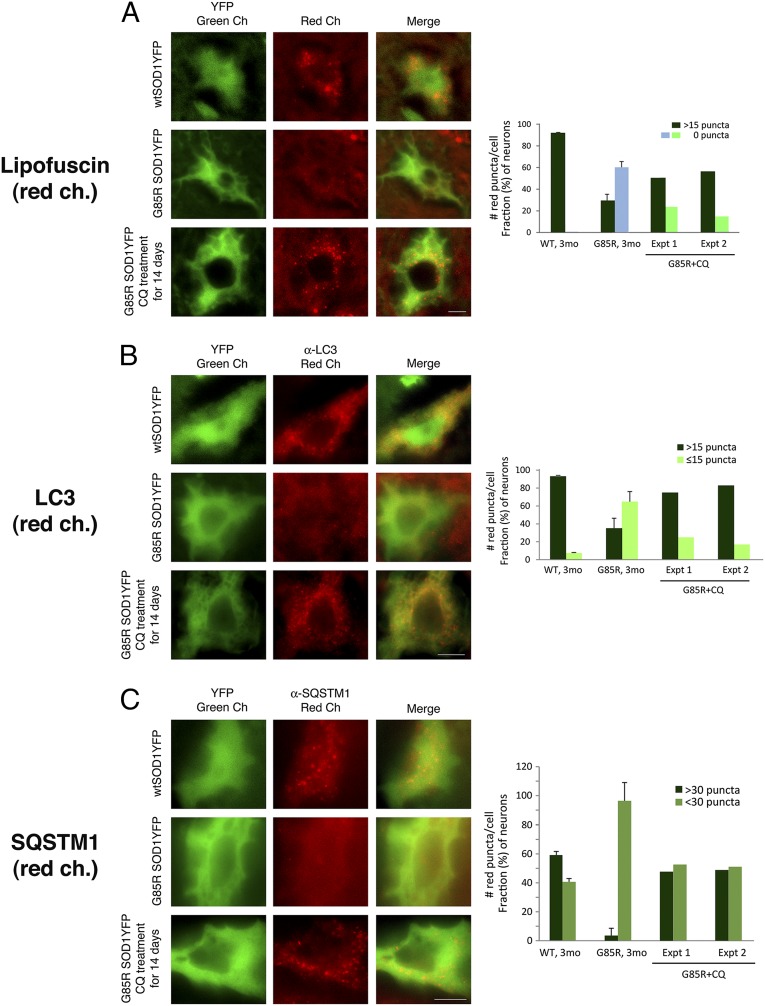
Fig. 3.
After treatment of G85R SOD1YFP mice with chloroquine, auto-fluorescent lipofuscin puncta, LC3, and SQSTM1 accumulate in motor neuron cell bodies. Two 3-mo-old G85R SOD1YFP mice were treated daily with chloroquine for 14 d as described in Materials and Methods. Spinal cord sections were prepared as described, and representative images are shown in the green channel for YFP and the red channel for (A) lipofuscin auto-fluorescence, (B) anti-LC3 immunofluorescence, and (C) anti-SQSTM1 immunofluorescence. In each set of panels, the top row shows WT SOD1YFP, the middle row shows G85R SOD1YFP, and the bottom row shows chloroquine-treated G85R SOD1YFP. For both lipofuscin and LC3, a substantial number of red puncta are present in the motor neuron cell body from the treated animal, suggesting that chloroquine, an inhibitor of lysosomal hydrolytic activity, had prevented the proteolysis of lipofuscin and LC3 components, thus permitting their accumulation. In the case of SQSTM1, a pattern of diffuse fluorescence in G85R SOD1YFP gives rise, upon treatment, to discrete puncta. The bar graphs on the right show the quantitation of the fraction of cells with different numbers of lipofuscin, LC3, or SQSTM1 puncta, respectively, in the two treated mice (two right-hand sets of bars, G85R+CQ, Exp. 1 and Exp. 2, for each mouse; >25 cells counted for each). The left-hand bars for WT and untreated G85R SOD1YFP animals are reproduced from Fig.1A in the case of lipofuscin, from Fig. 2A for LC3, and derived from Fig. 2B for SQSTM1. Note that both treated animals have an increased fraction of cells with the respective puncta. (Scale bars, 10 μm.)

