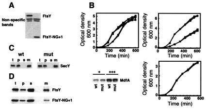
FIG. 2.
Characterization of E. coli EA101. (A) Extracts (20 μg of proteins) of E. coli EA101 (mut) and its parental strain, BW25113 (wild type [wt]), were analyzed by Western blotting with anti-FtsY antibodies. (B, top left panel) Growth of E. coli EA101 (solid symbols) and BW25113 (open symbols) was monitored in Luria-Bertani broth at 30°C (circles), 37°C (squares), and 42°C (triangles). (B, top right panel) Growth (37°C) of E. coli EA101 (solid symbols) and BW25113 (open symbols) was monitored in 2YT (rich medium; circles) and M9 (minimal medium; squares). (B, bottom left panel) E. coli EA101 (mut) and its parental strain, BW25113 (wt), moderately (+) or highly (+++) overexpressing MdfA-6His, were disrupted, and membranes (10 μg of proteins) were analyzed by Western blotting with horseradish peroxidase-conjugated INDIA HisProbe. (B, bottom right panel) The growth of the MdfA-overexpressing cells was monitored. (C) Membrane expression of SecY was analyzed by cell fractionation and Western blotting with anti-SecY antibodies obtained from H. Tokuda (University of Tokyo) (t, total; p, ultracentrifugation pellet; s, ultracentrifugation supernatant; m, membranes isolated by flotation). (D) Cellular distribution of FtsY-NG + 1. E. coli EA101 (mut) and its parental strain, BW25113 (wt), were fractionated, and the various fractions were analyzed by Western blotting with anti-FtsY antibodies.

