Fig. 5.
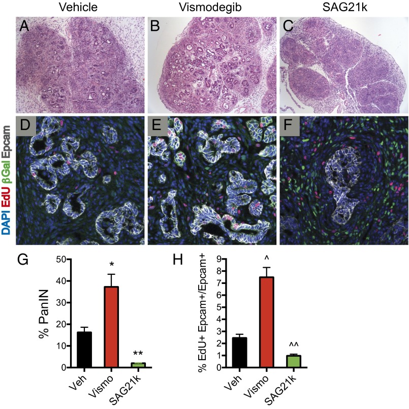
Hh response suppresses PanIN formation and proliferation. (A–C and G) H&E sections of pancreata from KICG mice given cerulein show PanIN lesions. Vismodegib-treated mice showed 37% of the pancreas occupied by PanIN lesions compared with 16% for vehicle-treated controls (*P = 0.016, n = 4 each). In contrast, SAG21k-treated mice showed only 2% of the pancreas occupied by PanIN lesions along with increased overall fibrosis (**P = 0.001, vehicle vs. SAG21k, n = 4). Error bars indicate SEM. (D–F and H) Representative confocal images of PanIN lesions in pancreata of KICG mice are shown. Vismodegib increases the percentage of proliferating PanIN cells (EdU+ Epcam+/Epcam+) to 7.5% compared with 2.5% for vehicle treatment (^P = 0.0004, n = 5 each). Conversely, SAG21k reduces proliferation percentage to 1.0% (^^P = 0.002, n = 5). Error bars indicate SEM.

