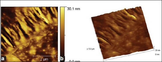
Figure 5.

(a) 2-Dimensional atomic force microscopy images of sucrose dependent biofilm formed by JKAS-CD2 cells. Following 12 h incubation, the microbial colonies adhered and seen embedded in the glucan produced.(b) 3-Dimensional AFM images of sucrose dependent biofilm formed by JKAS-CD2 cells. Following 12 h incubation, the microbial colonies adhered and seen embedded in the glucan produced
