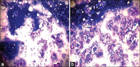
Figure 1.

Fine-needle aspiration cytology showing (a) Many anucleate squames and few nucleated benign squamous cells in a background containing neutrophils. Note the presence of benign ductal epithelial cells with myoepithelial component (May-Grünwald-Giemsa, ×200), (b) Anuclete squames, few nucleated benign squamous cells and neutrophils along with foreign-body type giant cell (arrow) (MGG, ×200)
