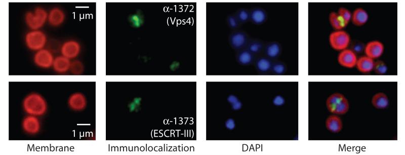
Fig. 2.
Localisation of (Top) Saci1372 (Vps4) and (bottom) Saci1373 (ESCRT-III). Representative images are shown. Images show the FM4-64X staining for membrane (red), DAPI staining for DNA (blue), antibody labelling of ESCRT-III or Vps4 (green) and merged images. Scale bar, 1 μm. Additional images are shown in figs. S6 and S7 and movie S1.

