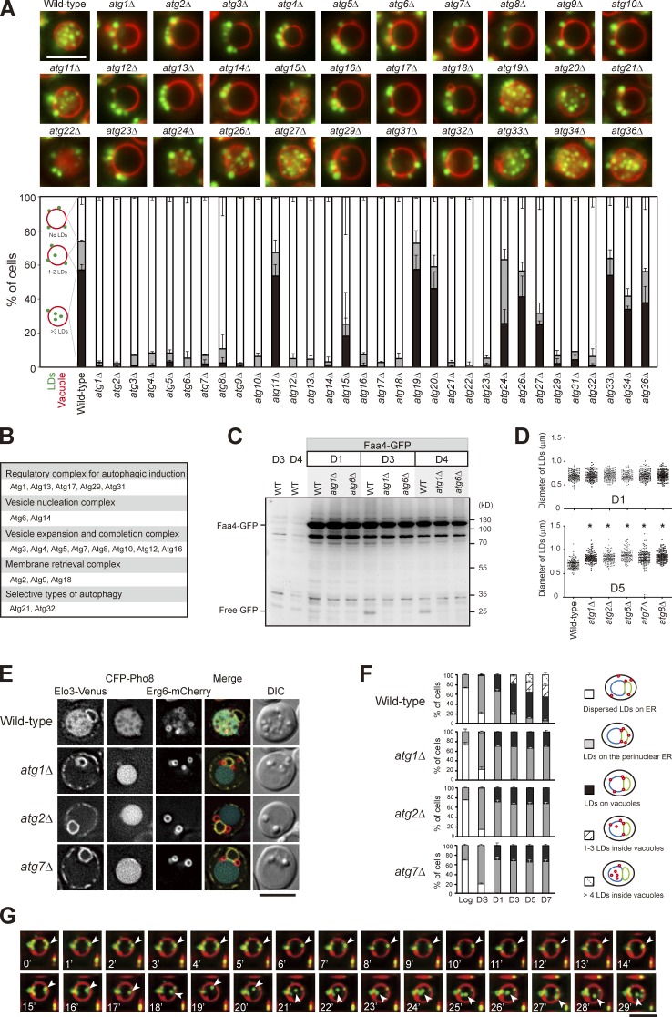
Figure 2.
Translocation of LDs into the vacuole lumen is mediated by a microautophagy mechanism. (A) Representative images of LDs and vacuoles in the indicated strains grown to D5. Quantification of localization based on the indicated patterns. Three independent data were plotted as mean ± SD. (B) Summary of A based on their roles in autophagy. (C) Wild type (WT) and various strains as indicated expressing Faa4-GFP were grown in SC medium to the indicated growth conditions. Cells were lysed, and the lysates were analyzed by immunoblotting with the anti-GFP antibody. (D) The comparison of LD diameter in the indicated strains grown to D1 and D5. The data shown are from one experiment (n = 100 for each cell type). *, P < 0.01. (E) Cells expressing the indicated proteins were grown in SC medium to D5 and imaged by fluorescence microscopy. DIC, differential interference contrast. (F) Quantification of data in E for the indicated localization patterns from three independent experiments, which were plotted as mean ± SD. (G) Wild-type cells stained with FM4-64 (vacuole) at DS and BODIPY (LDs) on D3 were subjected to time-lapse fluorescence microscopy. Images were taken every 1 min. Arrowheads denote an LD during its translocation into the vacuole lumen. Bars, 5 µm.

