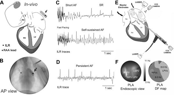
Figure 1.
In-vivo and ex-vivo model, protocols and mapping experiments. A) A diagram of a pacemaker with the lead in the RAA (light grey arrow) and ILR (dark grey arrow) close to the LA. B) Fluoroscopic view of the lead in the RAA (light grey arrow) and the ILR next to the LA (dark grey arrow). C) ILR traces at different stages of the persistent AF protocol. Rapid pacing appears at the beginning of the trace. D) A 6-sec long AF episode during AF that persisted for >7 days after turning off the fast pacing. E) Ex-vivo Epicardial and endocardial mapping setup includes synchronized dual CCD cameras as well as bipolar electrodes placed in the RA and the roof of the LA. Bipolar signals are also obtained from the pulmonary veins. F) Endoscopic view of the PLA (left) and DF map during AF.

