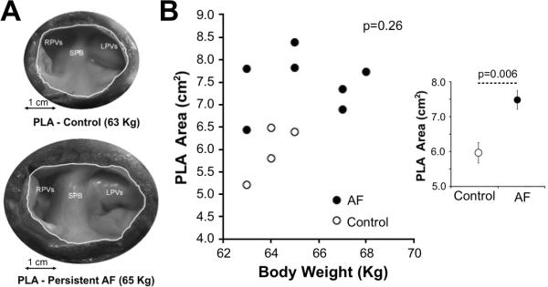
Figure 6.

Enlargement of the PLA area in sheep with AF. A) Macroscopic images of the endocardial side of the PLA area in a persistent AF sheep (bottom) and weight-matched control (top). B) No significant correlation between body weight and PLA area was observed (Spearman's test). Inset: On average, AF sheep show a significantly larger PLA area compared to weight-matched controls (Mann-Whitney U test).
