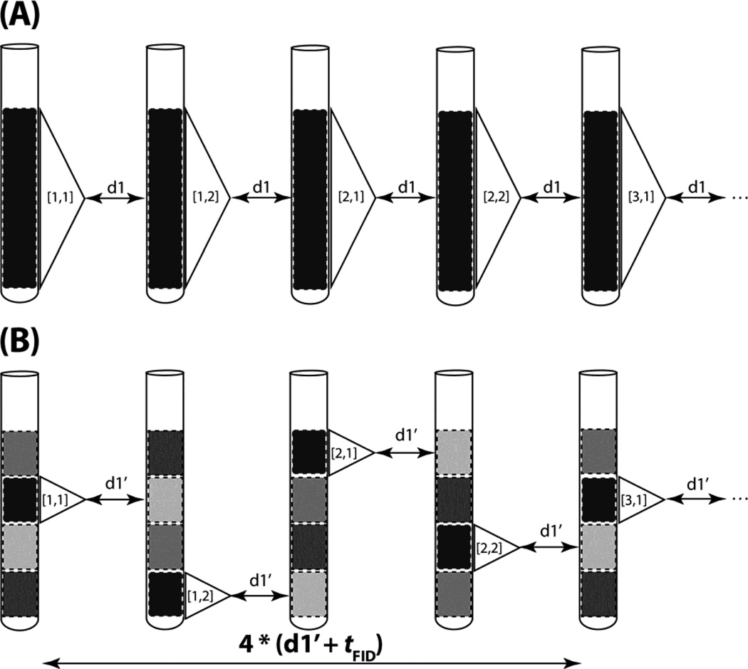
Figure 1.
Comparison of conventional with time-staggered data acquisition relying on spatial excitation. (A) Conventional data acquisition: the entire sample volume within the r.f. detection coil is utilized to record FIDs with a relaxation delay between scans denoted as d1. The excited region of the sample is depicted in black to indicate that a sizable steady state magnetization is present before excitation and the white dashed rectangle indicates the region for which the signal is detected. The FID is represented by a triangle and [N,P] within the triangle indicates the indirect dimension’s real data point N and its corresponding phase cycling step P. As an example, complex data acquisition (N = 1,2) is combined with a two step phase cycle (P = 1,2) cycle for axial peak suppression[15]. (B) Time-staggered data acquisition relying on spatial excitation of one slice of the entire volume at a time (highlighted with a white dashed rectangle). Considering tFID as the time required to detect an FID and d1’ as the relaxation delay between scans, the same steady state magnetization is present as in (A) if d1 + tFID = 4*(d1’ + tFID). The transitions from pale grey to grey, then to dark grey and eventually to black (second slice from the top) indicates progressing longitudinal relaxation in a slice while other slices are excited. As a result, NMR time domain data can be acquired more rapidly while ensuring that sufficiently long relaxation times between scans are employed.

