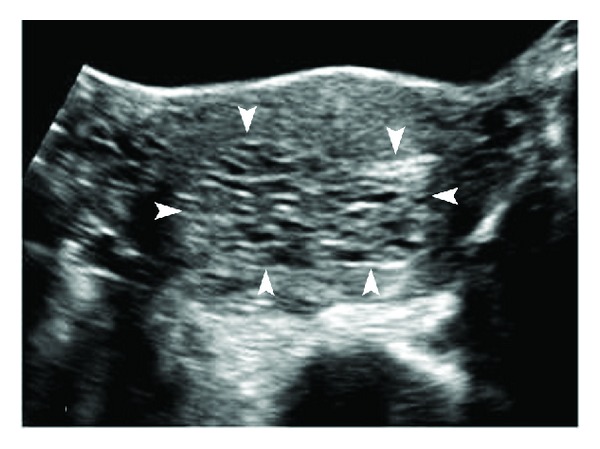
Figure 1.

Complete hydatiform mole. Transverse transabdominal sonography (TAS) image of the uterus shows distension of the uterine cavity by echogenic material with numerous small, irregular cystic spaces within (arrowheads). The normal hypoechoic myometrium can be seen stretched at the periphery. There is no identifiable fetal tissue.
