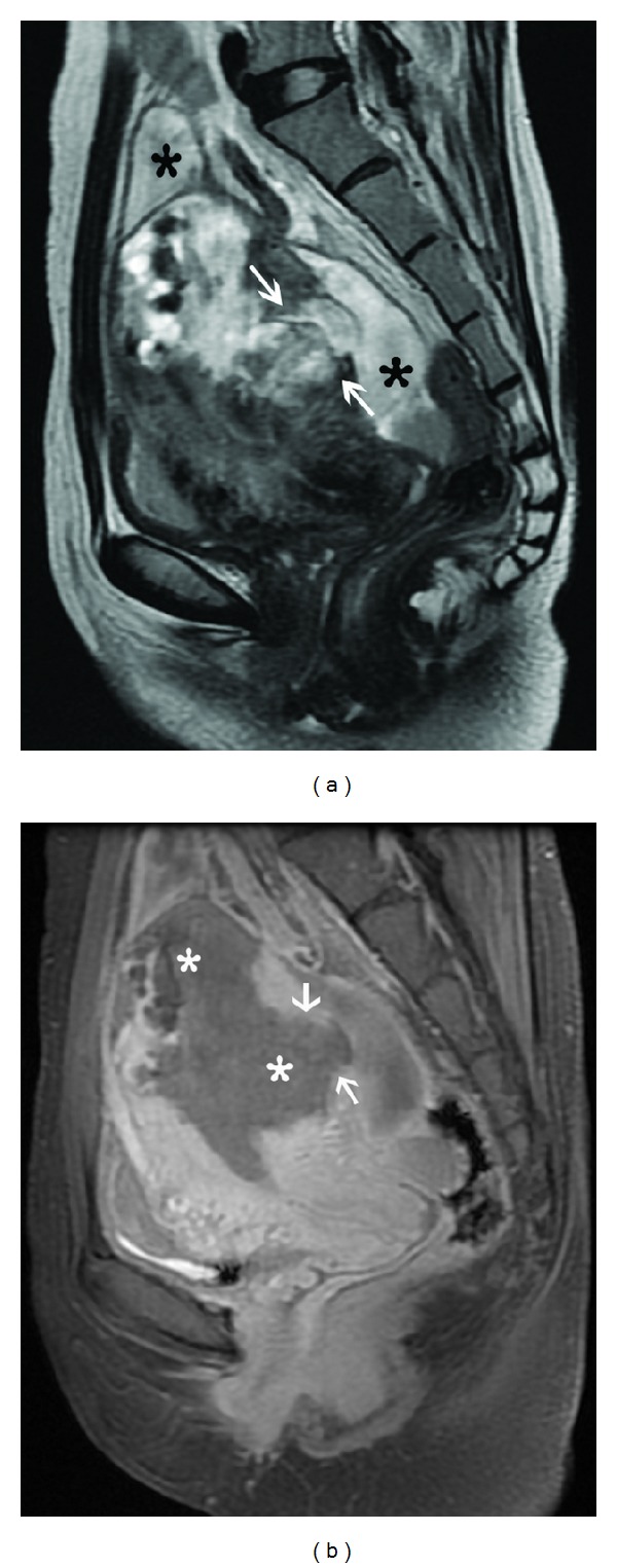
Figure 11.

Bulky choriocarcinoma with myometrial rupture. Sagittal T2-weighted image (a) shows an ill-defined heterogeneous mass in the uterine fundus and posterior corpus with full thickness myometrial penetration (arrows) and associated fluid collection (black asterisk) around the uterus. Sagittal contrast-enhanced fat-suppressed T1-weighted MR image (b) demonstrates the mass to be completely necrotic with a large myometrial perforation showing focal continuity (arrows) with the collection in the pouch of Douglas.
