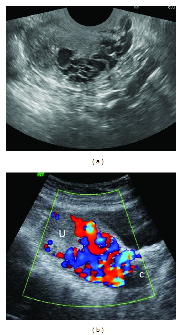
Figure 5.

Uterine vascular malformation presenting with recurrent vaginal bleeding following successful treatment of invasive mole. Sagittal TVS image (a) of the uterus reveals multiple tortuous, serpiginous anechoic spaces in the myometrium. Color Doppler TAS (b) reveals a mosaic pattern of color signal within the spaces. U = uterus; C = cervix. No evidence of any myometrial mass is seen to suggest a residual GTN.
