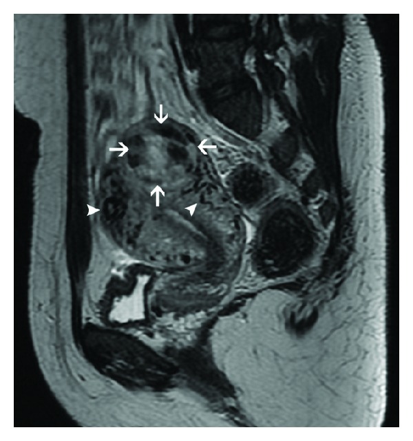
Figure 9.

Sagittal T2-weighted MR image in a patient with invasive mole demonstrates a heterogeneous, hyperintense uterine mass in the fundus with a myometrial epicenter (arrows). Tortuous flow voids (arrowheads) consistent with vessels are seen in the adjoining myometrium, indicative of tumor hypervascularity.
