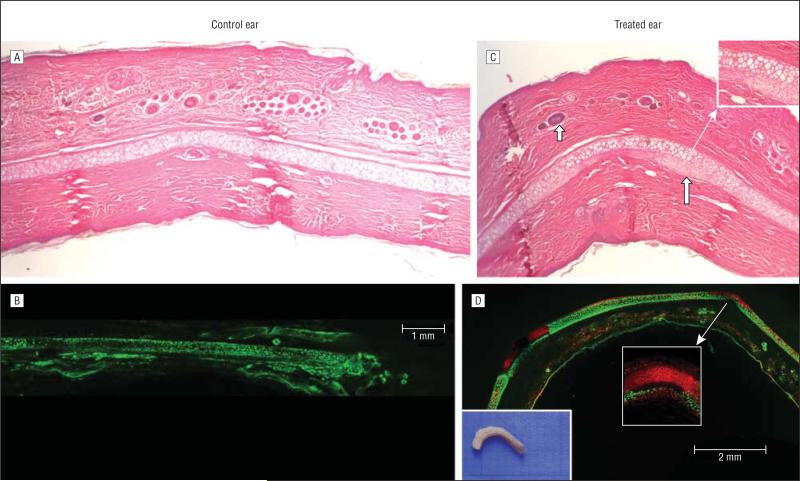
Figure 3.
Histologic findings. A, Control ear shows normal-appearing cartilage and epithelial lining. B, Live-dead fluorescent assay confocal microscopy shows viable chondrocytes (green) throughout the control specimen. C, Treated ear shows curved geometry with intact epithelial lining and adnexal structures (short blue arrow). Neochondrogenesis is noted along the undersurface of the cartilage in the periphery of perforated sites (long blue arrow and ×2 magnified inset). D, Live-dead fluorescent assay confocal microscopy shows limited zones of nonviable chondrocytes (red) around needle electrode sites, with live cells along the undersurface of the cartilage, presumably representing ingrowth of new chondrocytes (×10 magnified inset). The gross section is shown on the left.

