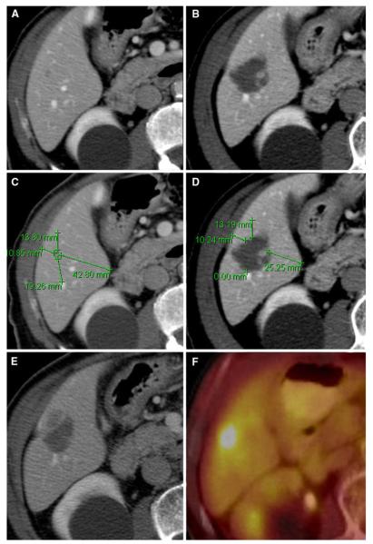
Fig. 5.
LTP of CLM after ablation from the site of the minimal margin (82-year-old male). A Preablation CT scan shows a small tumor located in the right hepatic lobe.
B Postablation CT shows the expected changes within the ablation zone. C Measuring the distance between the tumor edge and nearest reliable landmark in different directions on pre-CT. D Measuring the distance between the above same landmarks and ablation defect on post-CT; minimal margin was acquired to be 1–5 mm located at approximately 11 o’clock. E, F Five months later, CT and PET all showed local tumor progression at the site of this minimal margin

