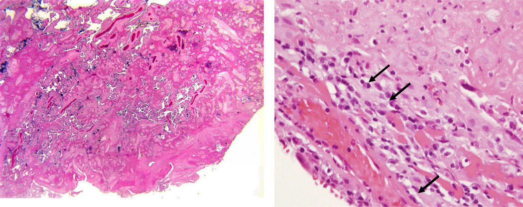
Figure 2.
Microscopic section of placenta from MPFD case #1. Note loss of normal villous architecture and encasement of remaining villi in pale-pink fibrin material (left). Area of chronic deciduitis with plasma cells (denoted by arrows) within large amount of fibrin deposition.This lesion is suggestive of maternal anti-fetal rejection (right).

