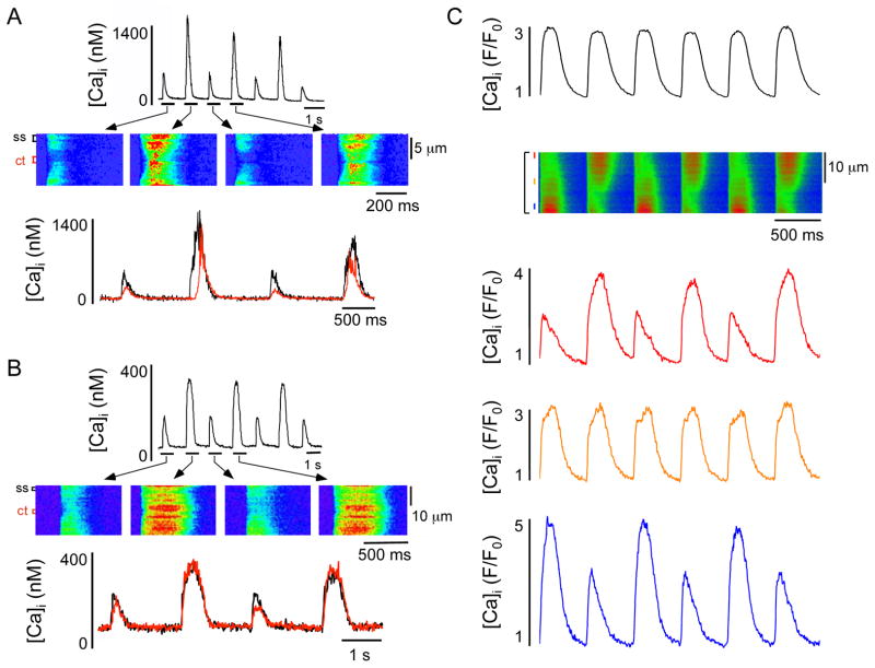
Figure 2. Cellular and subcellular Ca2+ alternans in cardiac myocytes.
A–B; Spatiotemporal characteristics of Ca2+ transients during alternans in an atrial (A) and ventricular (B) myocyte. From top: whole cell Ca2+ transients, transverse confocal line scan images and subcellular [Ca2+]i profiles recorded from subsarcolemmal (ss, black) and central (ct, red) regions of the myocyte. Panels A and B modified from Hüser et al. (10) with permission. C; Spatiotemporal characteristics of Ca2+ transients during alternans in an atrial myocyte where subcellular discordant or ‘out-of-phase’ alternans are present. The global [Ca2+]i profile suggests no Ca2+ alternans, however spatially restricted profiles identify subcellular regions with no alternans coexisting with regions alternating out-of-phase.

