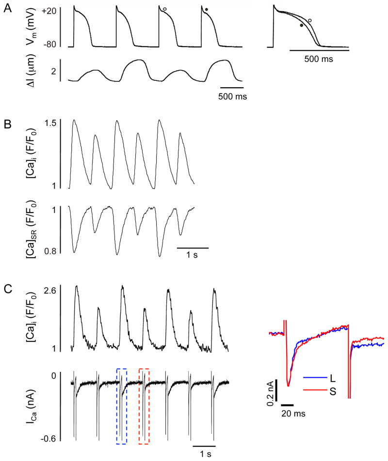
Figure 3. Electrical, mechanical and Ca2+ alternans in cardiac myocytes.
A; Simultaneous recordings of action potentials and cell shortening from a single ventricular myocyte revealing discordant electromechanical alternans. To the right, two action potentials recorded during successive small- (open circle) and large-amplitude (filled circle) shortenings are superimposed to illustrate the differences in duration and kinetics. Modified from Hüser et al. (10) with permission. B; Simultaneous recordings of cytosolic ([Ca2+]i; top) and intra-SR ([Ca2+]SR; bottom) Ca2+ alternans from a single ventricular myocyte. C; Simultaneous recordings of [Ca2+]i (top) and ICa (bottom) in voltage-clamped atrial myocytes. To the right, an overlay of ICa measured during a large-amplitude Ca2+ transient (L; blue trace) and a small-amplitude Ca2+ transient (S; red trace) shows that Ca2+ alternans are not accompanied by alternating peak ICa. Modified from Shrkyl et al. (67) with permission.

