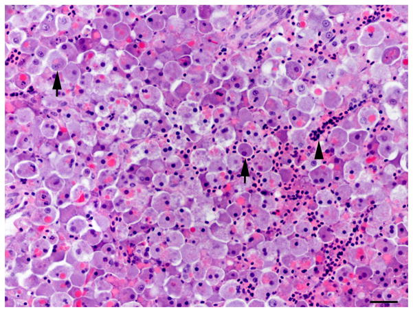
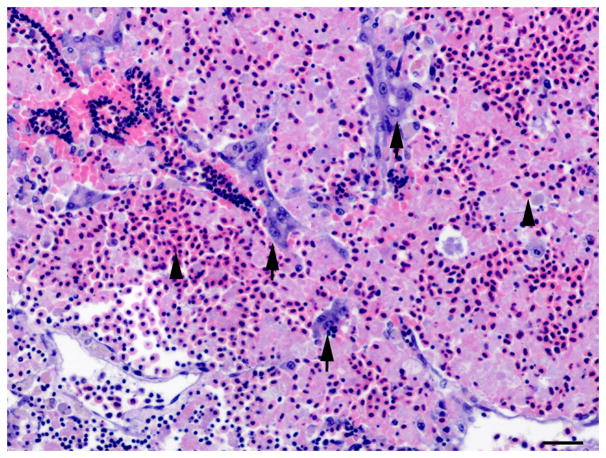
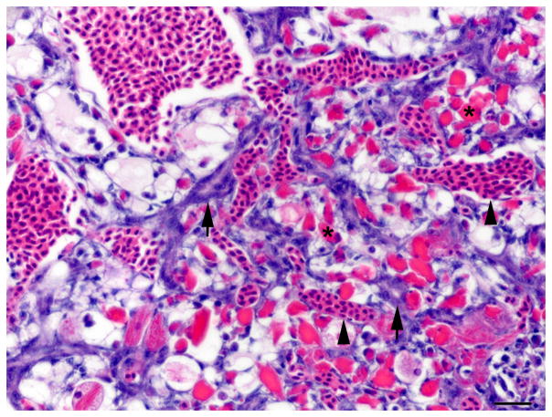
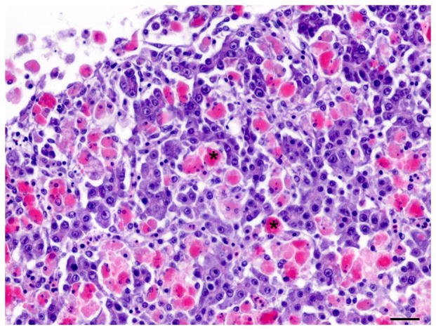
Fig. 1.
Histology of the regenerative liver response following microcystin LR (MCLR) exposure. (A) Severe diffuse peracute hepatic necrosis 14 h after MCLR exposure. The hepatic parenchyma is composed of dissociated rounded hepatocytes with pyknotic nuclei (arrow). HE. Bar, 30 μm. (B) Early hepatic regeneration 24 h after MCLR exposure. Basophilic oval cells (arrows) organized in clusters and tubules resembling the human ductular reaction are scattered within the necrotic hepatocellular parenchyma. HE. Bar, 30 μm. (C) Early hepatic regeneration 36 h after MCLR exposure. Tubules formed by basophilic oval cells often have a visible slit-like lumen (arrows) and are interconnected. Between the oval cells, numerous macrophages infiltrate the parenchyma (asterisks) and are filled with bright red cell debris due to ingestion of the hepatocytic RFP. HE. Bar, 30 μm. (D) Hepatic regeneration 48 h post exposure. The hepatic parenchyma is mostly composed of intermediate hepatobiliary cells and the infiltrate of macrophages (asterisk) is reduced. HE. Bar, 30 μm. Arrowheads in the figures indicate red blood cells.


