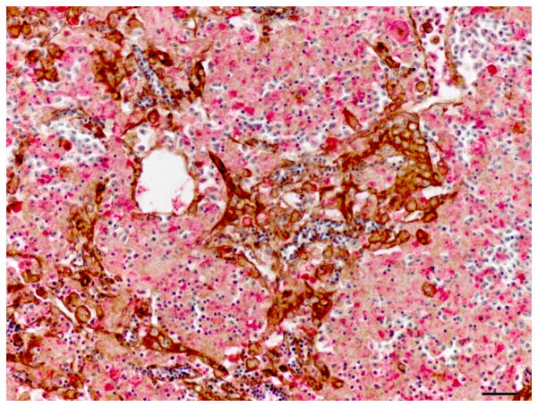
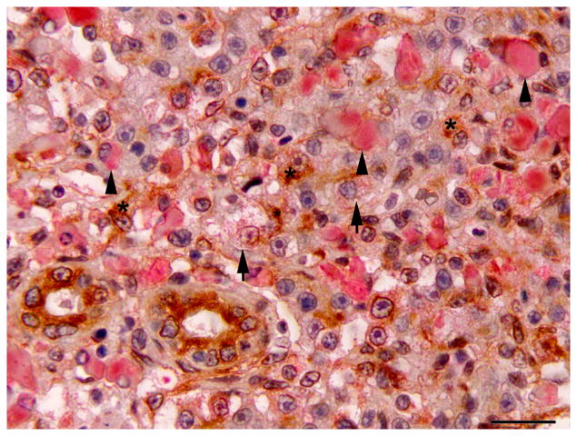
Fig. 2.
Hepatic regeneration following microcystin LR (MCLR) exposure; double immunolabelling for CK AE1/AE3 and red fluorescent protein (RFP). (A) Early hepatic regeneration 24 h after MCLR exposure. Oval cells (arrow) are strongly positive for CK (brown). Surrounding cell debris is variably immunoreactive for RFP (red). Double IHC for CK and RFP, haematoxylin counterstain. Bar, 30μm. (B) Hepatic regeneration 96 h post exposure to MCLR. The hepatic parenchyma is composed of intermediate hepatobiliary cells that are occasionally weakly positive for RFP (arrow). A small number of CK-positive oval cells are still present (asterisk). Cholangiocytes are also positive for CK. Macrophages display variable cytoplasmic expression of RFP (arrowheads). Double IHC for CK and RFP, haematoxylin counterstain. Bar, 20 μm.


