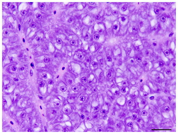
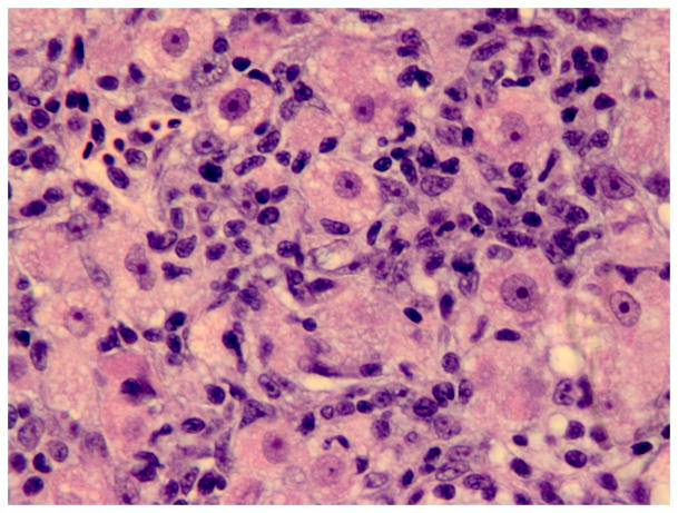
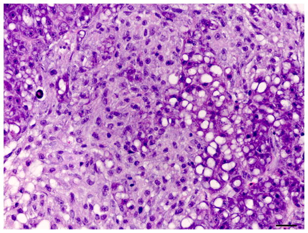
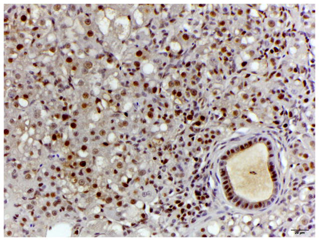
Fig. 3.
Histology of control liver and liver with oval cell hyperplasia. (A) Normal female medaka liver. HE. Bar, 20 μm. (B) Oval cell hyperplasia characterized by proliferation of small cells with a hyperchromatic elongated nuclei that separate the hepatocyte tubules and sometimes surround individual hepatocytes. HE. Bar, 20 μm. (C) Focus of oval cells differentiating towards hepatocytes. HE. Bar, 20 μm. (D) Liver, oval cell hyperplasia after DMN exposure. Oval cells, hepatocytes and cholangiocytes demonstrate nuclear immunoreactivity for PCNA of variable intensity. IHC. Bar, 50 μm.


