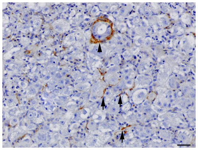
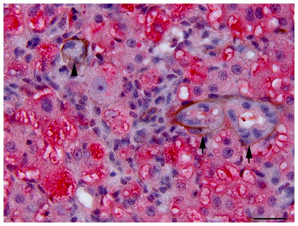
Fig. 5.
Single immunolabelling for muscle specific actin (MSA) and double immunolabelling for MSA and red fluorescent protein (RFP) in regenerating liver. (A) Oval cell hyperplasia after DMN exposure. Activated stellate cells (arrows) and myofibroblasts (arrow heads) are positive for MSA. IHC. Bar, 50 μm. (B) Oval cells and intermediate hepatobiliary cells admixed with hepatocytes after DMN exposure. Two immature bile ducts formed by RFP-negative intermediate hepatobiliary cells are surrounded by a thin rim of MSA-positive cells (arrows). Other RFP-negative intermediate hepatobiliary cells are surrounded by a rim of MSA-positive cells, but no duct lumen is present yet (arrowhead). Double IHC for MSA and RFP. Bar, 20 μm.


