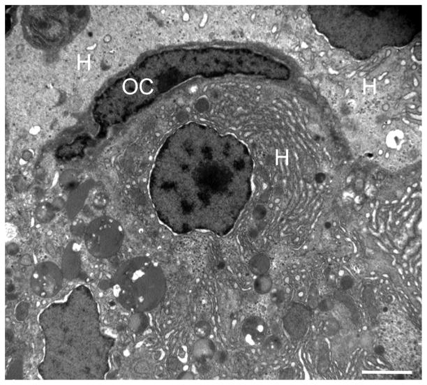
Fig. 6.
Transmission electron microscopy of the liver of a DMN-exposed medaka with oval cell hyperplasia. An oval cell (OC) is adjacent to three hepatocytes (H). Note the organelle-poor cytoplasm and lack of distinguishing morphological features of the OC. See Fig. 3b for the histological correlate. Bar, 2 μm.

